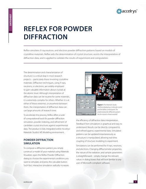
The online version of this article offers supplementary material ( ). Search in Google Scholar Supplementary Material Pandey, Close-Packed Structures, 2001st ed., University College, Cardiff Press, Wales University College, Cardiff, 1981. BIOVIA Materials Studio Reflex simulates X-ray, neutron, and electron powder diffraction patterns based on models of crystalline materials. These diffraction peaks were then indexed by the 4 indexing programs. Lotti, Cancrinite-Group Minerals at Non-Ambient Conditions: A Model of the Elastic Behavior and Structure Evolution, 2013. diffraction data obtained from X-ray, neutron, and electron radiation sources. The authors thank the two anonymous reviewers for their comments, which greatly improved the paper. Haishuang Zhao is grateful to the financial support from Carl-Zeiss-Stiftung. We gratefully acknowledge the German science foundation (DFG) for support in the large facility program project numbers INST144/435-1 FUGG and INST144/458-1 FUGG. Electron Back-Scatter Diffraction (EBSD) or Electron Back-Scatter Pattern (EBSP) is a powerful technique that captures electron diffraction patterns from crystals, con-stituents of material. Ternieden (Max-Planck-Institut für Kohlenforschung) for providing PXRD data in a time of need. Buhl for providing INT single crystals used in this work and to C. Using relative positions of the incorporated species in the natural cages as well as electron densities calculated by using only the framework of INT the positions of these species could be described. Through the comparison of simulated electron diffraction pattern to measured data the modeled framework could be confirmed. The possible combinations were further reduced by considering the amount of incorporated species. The average structure of INT was modeled considering only the combination of naturally existing (zeolitic) cages, restricted by the actual number of layers per unit cell. The average lattice and the stacking disorder along c axis could be confirmed by the reconstruction of three-dimensional ADT data.

The hexagonal lattice parameters with a=1266.3(2) pm and c=1586(1) pm and the unit cell setting were determined by Pawley fits. The comparison of the intensity ratios of PXRD data with a SCXRD measurement indicates the formation of a comparable phase with the typical strong stacking disorder. The formation of the INT phase was proved by both PXRD and TGA analysis and its stoichiometric composition was found to be |Na 6.95(1)(CO 3) 0.48(2) (H 2O) 6.18(6)| 6. Powder samples of the intermediate phase between sodalite and cancrinite (INT) have been synthesized hydrothermally.


 0 kommentar(er)
